Bones In Your Leg Diagram / 16 Bones In The Leg Ideas Anatomy Leg Anatomy Leg Bones / Pain in the large bones and muscles of the upper leg can be caused by a wide range of ailments.
byAdmin-
0
Bones In Your Leg Diagram / 16 Bones In The Leg Ideas Anatomy Leg Anatomy Leg Bones / Pain in the large bones and muscles of the upper leg can be caused by a wide range of ailments.. Femur (2 bones) patella or kneecap (2 bones) tibia (2 bones) fibula (2 bones) foot (52 bones in total, 26 per foot) tarsus/tarsals. The bones of the appendicular skeleton provide support and flexibility at the joints and anchor the muscles that move the limbs. They are numbered from one to five, starting from the medial (inner) side of the foot. Home » unlabelled » bones in leg diagram / your leg bones are very large and strong to help support the weight of your body. Tendons connect the knee bones to the leg muscles that move the knee.
Let's review all of these bones one last time. The knee joint is the largest joint in the body and is primarily a hinge joint, although some sliding and rotation occur. The pelvic region is the area between the trunk — or main body — and the lower extremities, or legs. The femur bone is the toughest and longest bone in the body, employing the location of the lower limb, between the hip and knee joints. This allows weight to be distributed either anteriorly or posteriorly throughout the foot.
Bones Of The Lower Limb Anatomy And Physiology I from s3-us-west-2.amazonaws.com The bones of the skeletal system act as attachment points for the skeletal muscles of the body. This allows weight to be distributed either anteriorly or posteriorly throughout the foot. The bones of the foot are organized into the tarsal bones, metatarsal bones, and phalanges. Ankle & lower leg anatomy. This keeps the bones together, giving a high ankle sprain time to heal. The bones together make up the hip. The second metatarsal bone is the longest. The knee joint is the largest joint in the body and is primarily a hinge joint, although some sliding and rotation occur.
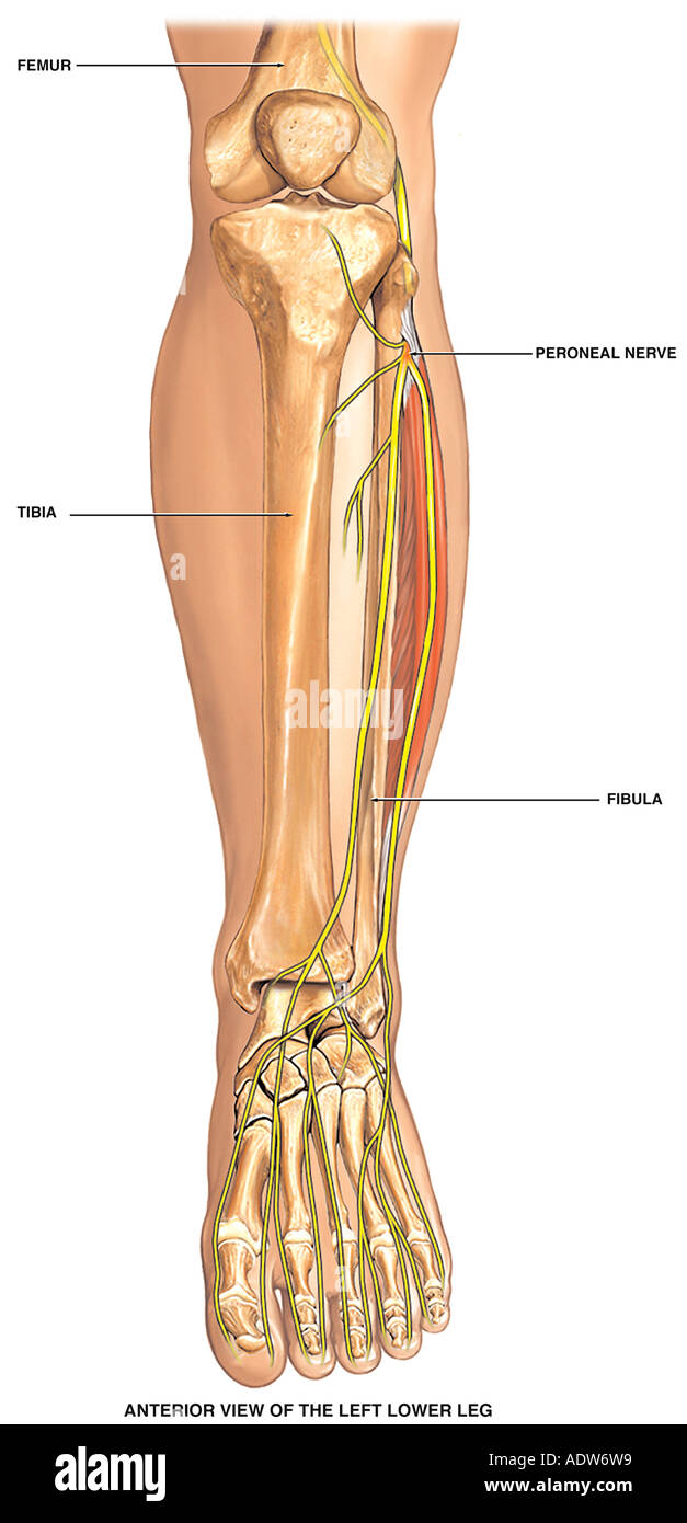
The foot begins at the lower end of the tibia and fibula, the two bones of the lower leg.
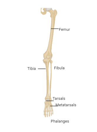
The bones of the hip include the femur, the ilium, the ischium, and the pubis. These muscles work together to produce movements such as standing, walking, running, and jumping. The smaller bone that runs alongside the tibia (fibula) and the kneecap (patella) are the other bones that make the knee joint. The bones of the leg are the femur, tibia, fibula and patella.the foot bones shown in this diagram are the talus, navicular, cuneiform, cuboid, metatarsals and calcaneus. The five metatarsals are the long bones that link the tarsal bones to the toes, seen in yellow in the diagram below. This allows weight to be distributed either anteriorly or posteriorly throughout the foot. There are a total of 60 bones in the legs. Tibia and fibula the tibia and fibula are two long bones that run parallel to each other, forming the scaffold of the leg and providing attachment points for many muscles. The talus the weight of your body is transferred from the tiba to the talus. The femur bone is the toughest and longest bone in the body, employing the location of the lower limb, between the hip and knee joints. Pain in the large bones and muscles of the upper leg can be caused by a wide range of ailments. Your hamstring is directly connected to your ischium bones, and any tear or damage to your hamstring can result in sit bone pain. The bones of the appendicular skeleton provide support and flexibility at the joints and anchor the muscles that move the limbs.
More specifically, this beautifully illustrated anatomy chart includes neck and shoulders, multiple views of the back and spine, and frontal views of each muscular extremity of the human body. The lower leg is comprised of two bones, the tibia and the smaller fibula. Its lower end helps create the knee joint. Many muscles that move the trunk and legs, such as our abdominal muscles, attach to the hip bones. The lower limb contains 30 bones.
Lower Leg Anatomy High Resolution Stock Photography And Images Alamy from c8.alamy.com The femur figure is so exclusive that it makes the bone satisfactory for bolstering the numerous muscular and ligamentous connection s within this zone, in addition to maximally stretching the limb during ambulation. The pelvic region is the area between the trunk — or main body — and the lower extremities, or legs. Tendons connect the knee bones to the leg muscles that move the knee. The second metatarsal bone is the longest. The bones of the leg are the femur, tibia, fibula and patella.the foot bones shown in this diagram are the talus, navicular, cuneiform, cuboid, metatarsals and calcaneus. There are a total of 60 bones in the legs. Almost every skeletal muscle works by pulling two or more bones either closer together or further apart. The talocrual joint is made up of three main bones.
This keeps the bones together, giving a high ankle sprain time to heal.
In addition, the broad hip bones provide protection to the delicate internal organs of the pelvis, such as the intestines, urinary bladder, and uterus. This muscle runs along the outside of the back of your thigh and attaches to the top of the fibula (the smaller of the two bones of your lower leg). Also called the shin bone, the tibia is the longer of the two bones in the. The smaller bone that runs alongside the tibia (fibula) and the kneecap (patella) are the other bones that make the knee joint. Depending on the cause, leg lumps may be single or while these conditions are rare, both benign and malignant tumors of the skin, soft tissues, or bones can sometimes feel. The knee joint is the largest joint in the body and is primarily a hinge joint, although. The thigh bone, or femur, is the large upper leg bone that connects the lower leg bones (knee joint) to the pelvic bone (hip joint). The five metatarsals are the long bones that link the tarsal bones to the toes, seen in yellow in the diagram below. The femur figure is so exclusive that it makes the bone satisfactory for bolstering the numerous muscular and ligamentous connection s within this zone, in addition to maximally stretching the limb during ambulation. The hip itself is a ball and socket joint, much like the shoulder.the structures necessary to create this joint are the socket, the joint capsule, muscle, ligaments, and the neck. The bones of your leg and foot helped give you the ability to score that field goal. Distal to the ankle is the foot. Pain in the large bones and muscles of the upper leg can be caused by a wide range of ailments.
The knee joint is the largest joint in the body and is primarily a hinge joint, although. This allows weight to be distributed either anteriorly or posteriorly throughout the foot. The lower leg extends from the knee to the ankle. Femur (2 bones) patella or kneecap (2 bones) tibia (2 bones) fibula (2 bones) foot (52 bones in total, 26 per foot) tarsus/tarsals. The thigh bone, or femur, is the large upper leg bone that connects the lower leg bones (knee joint) to the pelvic bone (hip joint).
Given Diagram Shows The Bone Of The Left Human Hindlimb Class 11 Biology Cbse from www.vedantu.com The bones of the leg are the femur, tibia, fibula and patella.the foot bones shown in this diagram are the talus, navicular, cuneiform, cuboid, metatarsals and calcaneus. They are numbered from one to five, starting from the medial (inner) side of the foot. The talus the weight of your body is transferred from the tiba to the talus. Almost every skeletal muscle works by pulling two or more bones either closer together or further apart. The bones of the skeletal system act as attachment points for the skeletal muscles of the body. The bones of the hip include the femur, the ilium, the ischium, and the pubis. Femur (2 bones) patella or kneecap (2 bones) tibia (2 bones) fibula (2 bones) foot (52 bones in total, 26 per foot) tarsus/tarsals. The bones of the appendicular skeleton provide support and flexibility at the joints and anchor the muscles that move the limbs.
The femur, or thighbone, is the longest and largest bone in the human body.
In addition, the broad hip bones provide protection to the delicate internal organs of the pelvis, such as the intestines, urinary bladder, and uterus. The bones of the foot are organized into the tarsal bones, metatarsal bones, and phalanges. The bones of the skeletal system act as attachment points for the skeletal muscles of the body. He leg's main function in the human is for locomotion and support of the rest of the body. More specifically, this beautifully illustrated anatomy chart includes neck and shoulders, multiple views of the back and spine, and frontal views of each muscular extremity of the human body. The pubis, ischium, and ilium together constitute the pelvis while the thigh bone is the femur. Femur (2 bones) patella or kneecap (2 bones) tibia (2 bones) fibula (2 bones) foot (52 bones in total, 26 per foot) tarsus/tarsals. The hip itself is a ball and socket joint, much like the shoulder.the structures necessary to create this joint are the socket, the joint capsule, muscle, ligaments, and the neck. The smaller bone that runs alongside the tibia (fibula) and the kneecap (patella) are the other bones that make the knee joint. This muscle runs along the outside of the back of your thigh and attaches to the top of the fibula (the smaller of the two bones of your lower leg). The lower leg extends from the knee to the ankle. The medial, larger bone of the lower leg. Distal to the ankle is the foot.
These muscles work together to produce movements such as standing, walking, running, and jumping bones in leg diagram. Tibia and fibula the tibia and fibula are two long bones that run parallel to each other, forming the scaffold of the leg and providing attachment points for many muscles.